
Cardiovascular disease rarely develops as an isolated problem in one artery or one organ. Atherosclerosis and vascular calcification can reflect a broader systemic process influenced by age, genetics, tobacco exposure, blood pressure, cholesterol, diabetes, kidney disease, nutrition, physical activity, sleep and inflammation. Calcification found in one vascular bed may be a signal to examine overall cardiovascular risk rather than focusing only on the location where it was discovered.
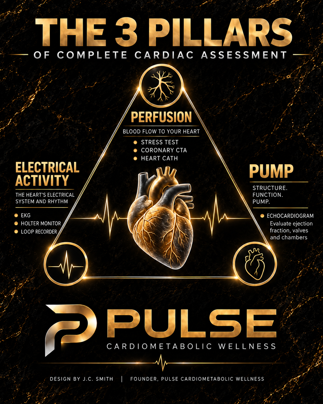
That is why a whole-person approach matters. Testing can identify disease, but prevention also requires attention to the factors that allow disease to progress. A useful cardiac assessment considers three clinical pillars: the heart’s structure and pumping function, its electrical activity and blood flow to the heart muscle. The tests within those pillars should be selected according to symptoms, history, physical findings and overall risk. More testing is not automatically better; the right test is the one that answers a meaningful clinical question.
Begin with a cardiovascular baseline
For someone who wants to establish a baseline, the first step is a careful history and risk assessment. This includes blood pressure, lipid profile, glucose or A1C, smoking history, family history of premature cardiovascular disease, weight and waist trends, exercise tolerance, sleep, diet, medications and current symptoms. A physical examination can identify findings such as a heart murmur, irregular rhythm, edema, abnormal pulses or signs of vascular disease.
An echocardiogram and electrocardiogram may be appropriate parts of that baseline when the history, examination, symptoms or risk profile justify them. They are not universal screening tests for every asymptomatic adult. This distinction protects patients from unnecessary testing while still allowing early evaluation when there is a clinical reason to look deeper.
Pillar one: structure, function and pump
A transthoracic echocardiogram uses ultrasound to evaluate the heart’s anatomy and performance. It can assess chamber size, wall thickness, heart muscle motion, ejection fraction, valve function, pressure estimates and the tissue surrounding the heart. In practical terms, it answers three questions: Is the heart built normally, does it relax and contract normally, and do the valves move blood in the correct direction? The American Heart Association notes that echocardiography provides information about both cardiac structure and function.
An echocardiogram is particularly valuable when a patient has a murmur, shortness of breath, swelling, abnormal ECG findings, suspected cardiomyopathy, known valve disease, heart failure symptoms or a meaningful change in functional capacity. When an abnormality is found, the study becomes a reference point for future comparison. The timing of repeat imaging depends on the condition and whether symptoms change.
Pillar two: electrical activity
A standard 12-lead electrocardiogram, commonly called an ECG or EKG, records the electrical activity of the heart at a single moment. It can show heart rate, rhythm, conduction patterns, prior injury patterns and clues to chamber enlargement or other cardiac conditions. It is fast, painless and useful when a patient reports palpitations, dizziness, fainting, chest discomfort, unexplained fatigue or an irregular pulse.
A normal office ECG does not exclude an intermittent rhythm problem. When symptoms come and go, ambulatory monitoring may be more useful. A Holter monitor typically records continuously for a shorter period, while extended patch monitors or event monitors can capture less frequent episodes. The goal is to connect a symptom with the heart rhythm at the same time. Monitoring can identify atrial fibrillation, significant bradycardia, supraventricular tachycardia, ventricular arrhythmias or conduction disease, but it should be matched to the frequency and seriousness of the symptoms.
Pillar three: perfusion and coronary disease
Perfusion refers to blood flow reaching the heart muscle. Symptoms are central to deciding how to evaluate it. Classic stable angina is often described as pressure or discomfort behind the breastbone that appears with exertion or emotional stress and improves with rest. Other warning signs can include a new decline in exercise capacity, exertional shortness of breath, jaw or arm discomfort, nausea, sweating or unusual fatigue. Symptoms may be less typical in women, older adults and people with diabetes.
Several tests answer different questions:
- A coronary artery calcium score measures calcified plaque and can refine preventive decisions when risk or the need for statin therapy remains uncertain. It is generally not the preferred test for active chest pain.
- An exercise treadmill test evaluates symptoms, rhythm, blood pressure response and ECG changes during exertion in appropriately selected patients.
- Stress echocardiography or nuclear myocardial perfusion imaging evaluates whether exertion or medication produces evidence of reduced blood flow.
- Coronary CT angiography shows coronary plaque and narrowing. In selected cases, fractional flow reserve derived from CT data can estimate whether a narrowing is likely to limit blood flow.
If noninvasive testing shows significant ischemia, if symptoms remain concerning despite treatment or if the clinical risk is high, invasive coronary angiography during left heart catheterization may be appropriate. Catheterization can define the coronary anatomy and measure pressures, and it can allow angioplasty or stent placement when indicated. It is not the automatic next step after every abnormal screening result; the decision depends on symptom burden, anatomy, ischemic risk, kidney function, bleeding risk and the expected benefit of intervention.
The systemic view of cardiovascular risk
A test result is only one part of the picture. Coronary calcification represents established atherosclerotic plaque, but the same biology may affect arteries supplying the brain, kidneys, abdomen and legs. That systemic view should prompt a review of modifiable risk factors rather than fear or indiscriminate scanning. The highest-value plan usually addresses tobacco exposure, blood pressure, atherogenic cholesterol, glucose control, physical activity, nutrition, sleep and medication adherence. Genetics and age cannot be changed, but they can influence how early and how aggressively modifiable risks should be treated.
At Pulse Cardiometabolic Wellness, my focus is to connect testing to a practical plan. That may include reviewing symptoms and family history, coordinating appropriate cardiac or vascular studies, improving metabolic health, supporting sustainable weight management and monitoring the measures that influence long-term risk. The purpose of establishing a baseline is not to order every available test. It is to identify what is normal, recognize what requires surveillance and act early on the risks that can be changed.
When symptoms require urgent care
New or severe chest pressure, symptoms at rest, shortness of breath, fainting, sweating, nausea or pain spreading to the arm, jaw, back or shoulder can represent an emergency. Call 911 rather than waiting for an outpatient appointment. A baseline assessment is preventive care; it is not a substitute for emergency evaluation.
Clinical references
American Heart Association: Echocardiogram
American Heart Association: Electrocardiogram
American Heart Association: Coronary Artery Calcium Test
American Heart Association: Exercise Stress Test
American Heart Association: Cardiac Computed Tomography Angiography
This article is for general educational purposes only and does not constitute medical advice or establish a provider-patient relationship. Testing decisions require an individualized evaluation by a qualified healthcare professional. New or severe chest pressure, symptoms at rest, shortness of breath, fainting, sweating, nausea or pain spreading to the arm, jaw, back or shoulder may represent an emergency; call 911.